Hypoparathyroidism May Lead to Rare Neurological Disease, Case Study Suggests
Written by |


Hypoparathyroidism may lead to the development of a rare neurological disorder manifesting with seizures, a case study reveals.
The study, “A Case of Seizure Revealing Fahr’s Syndrome with Primary Hypoparathyroidism,” was published in the American Journal of Case Reports.
The patient, a 52-year-old man, was admitted to a hospital because of a sudden seizure, during which his arms and legs were slightly tilted for about 10 minutes (generalized tonic-clonic type seizure).
The man did not have a history of seizures, head trauma, central nervous system infection, stroke, hypertension, diabetes, thyroid disease, or autoimmune disease. He also reported no family history of epilepsy.
During neurological examination, the patient was clearly aware, without signs of cognitive impairment or cranial nerve abnormalities.
Laboratory analysis revealed reduced levels of calcium and slightly higher levels of phosphate in the blood. The patient was also secreting slightly more calcium (0.17 mg/dL) than normal (less than 0.14 mg/dL), according to urine analysis results.
Further analysis showed that he had much lower levels of parathyroid hormone (PTH, 3.5 pg/dL) in the blood compared to normal values (15–65 pg/dL).
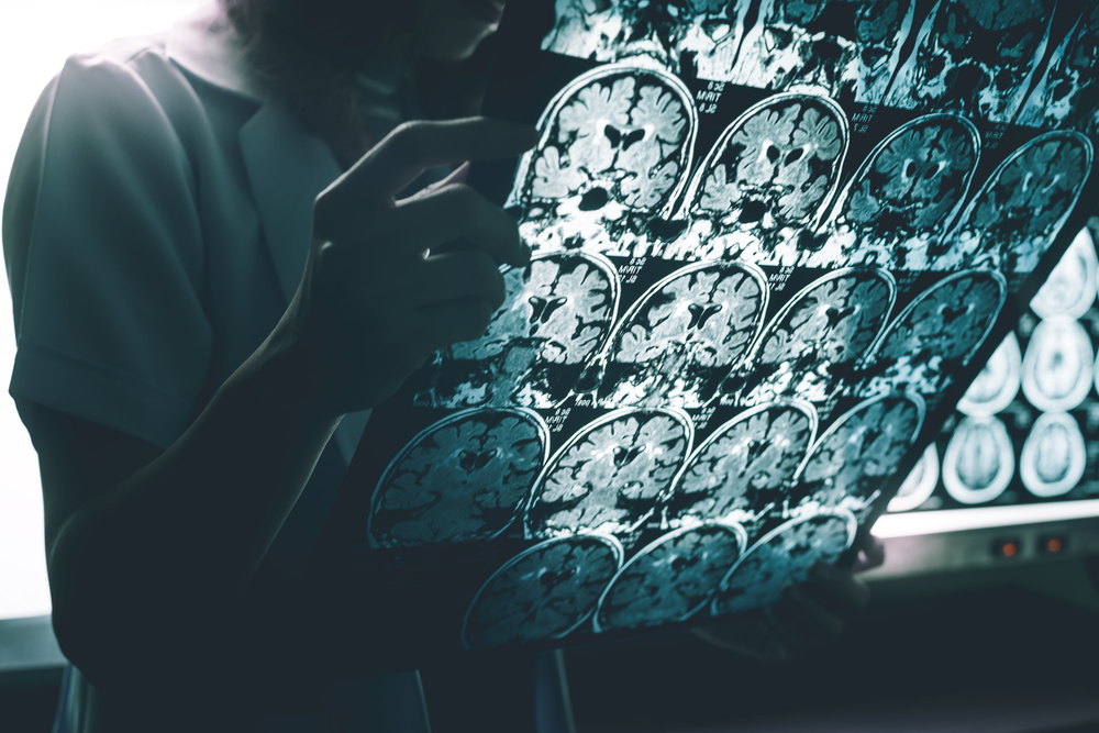
Ultrasound evaluation of his thyroid did not show any significant structural alterations of the gland or surrounding tissues. However, brain images by computed tomography (CT) revealed that the patient had calcifications within the structures of the basal ganglia (a region deep in the center of the brain, involved in coordination of movement).
Evaluation of brain activity by electroencephalography (EEG) did not reveal any signs of active seizures or activity changes.
Reduced parathyroid hormone levels suggested hypocalcemia due to hypoparathyroidism. “Primary hypoparathyroidism was diagnosed because there were no indications of secondary causes such as a history of congenital anomalies, thyroid surgery [or] history of neck radiation,” researchers said.
Based on his CT results, the patient was diagnosed with Fahr’s syndrome caused by hypoparathyroidism.
Fahr’s syndrome is a rare neurological disorder characterized by the abnormal accumulation of calcified deposits in the basal ganglia. It often manifests with deterioration of motor function, dementia, seizures, headache, stiffness of the limbs, and involuntary writhing movements.
To correct calcium levels, the patient received calcium gluconate intravenously for 24 hours. He also started a regime of oral medication, including calcium carbonate three times a day and Rayaldee (calcifediol) and hydrochlorothiazide (sold under the name Microzide, among others) once a day.
To prevent seizures, he was treated with topiramate (sold as Topamax or Qudexy XR) twice a day.
“Although there are many specific hypotheses, the causes of abnormal calcification in the brain are related to parathyroid dysfunction or other causes of calcium metabolism overall,” researchers said.
“Although rare, Fahr’s syndrome should be suspected if there is symmetrical and abnormal calcification of both basal ganglia, visible via brain CT, of patients with seizures or loss of consciousness. Moreover, thyroid and parathyroid function should be tested along with the calcium concentration in the blood. … We believe that confirming brain imaging in hypoparathyroidism patients with all neurological symptoms will help to improve the prognosis,” they stated.